
The 4 Natural Curves of Your Spine
- Dr. Jill Evans

- Aug 17, 2021
- 4 min read

One of the biggest misconceptions among chiropractic patients is that a straight spine is a healthy spine. Any chiropractor knows this is far from the truth. The reality is that an ideal spine is one with healthy curvature – specifically an “S” curve from top to bottom. It’s this curve that enables the spine to function properly as the central core to our balance and stability. It also helps with:
Better movement
Lesser aches and pain
Stronger posture
Healthier life
Better sport performance
Lesser restricted motion
More Flexible
Protection of the spinal cord and nerve roots
Kempsville Chiropractic has done a wonderful job of breaking down the spine very simply.
The cervical curve, better known as your neck, should also be slightly curved inward to complete the S-shape. Leaning too far forward leads to a hunched-over look while leaning too far back scrunches your neck. This is where the straight line comes in: When you look at the side view of someone standing with proper posture, a straight line should run from their ear to their ankle. If your line is uneven, the cervical curve may be to blame.
The cervical spine, or neck, consists of the first 7 vertebrae labeled C1 through C7. The nerves running through the cervical spine supply the head, neck, and shoulder areas. Science has been able to directly correlate various nerves and their function to individual vertebrae in the spine.
The nerves running through the upper half of your neck supply your brain, intracranial blood vessels, eyes, lacrimal gland, parotid gland, pituitary gland, scalp, sinuses, neck and facial muscles, hearing, and more.
The lower half of your neck supplies your neck and shoulder muscles, elbows, arms, wrists, hands, esophagus, tonsils, thyroid gland, and even some facets of your heart, lungs, and chest.
As you can see, the neck controls the nerve supply to numerous and important parts of the body. If there are subluxations, or misalignments, in your neck (cervical spine) they can cause many problems if left uncorrected. Chiropractors are able to make adjustments to subluxations (misalignments) in the neck to help patients deal with problems. Many symptoms in our patients have been linked directly to subluxations in the cervical spine.
The thoracic curve is named for the thorax, the area between the neck and the abs. It's the part of the spine responsible for hunching and slouching. The thoracic spine consists of 12 vertebrae (spinal bones) located in the mid to upper back and is labeled T1 – T12. It is located just below the cervical spine (neck) and above the lumbar spine (lower back). In addition to providing protection to the spinal cord, the thoracic vertebrae are what your ribs connect to. Unlike the cervical and lumbar spines with both curves inward (toward the body – lordosis), the thoracic spine curves outward (away from the body – kyphosis) which allows you to be able to bend over and touch your toes.
The nerves that exit through the thoracic spine control the function of many major organs in the body. The nerves exiting through the T1 to T4 vertebrae are associated with the functions of the heart, lungs, esophagus, breast, larynx, trachea, parts of the arms, and muscles of the upper body. T5 through T10 nerves correspond with your gallbladder, liver, stomach, diaphragm, pancreas, spleen, kidneys, small intestine, adrenal and suprarenal glands, and appendix. While the nerves exiting the T11 and T12 vertebrae are related to your small intestines, colon, uterus, lymph circulation, solar plexus, and muscles in your mid to upper body.
The lumbar curve connects your abs to your lower body. These vertebrae should be slightly curled inward. We lose this natural form when we lean back in our chairs and slide our bottoms forward. You can recover the curve by sitting back in your chair with hips at a 90-degree angle to the back of the chair and your shoulders pushed back. You're working those postural muscles!
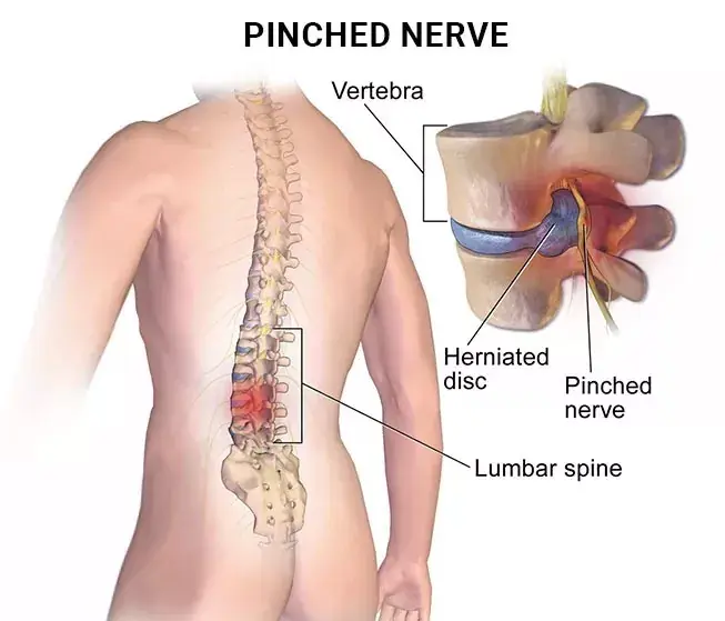
The lumbar spine consists of five vertebrae (spinal bones) labeled L1 to L5, which are located in the lower back below the thoracic spine. The lumbar spine contains the largest and strongest vertebrae in the spine, and in addition to permitting movement, it supports the majority of the weight of the upper body. The large nerves that exit the lumbar spine control and coordinate some of the largest and strongest muscles in the body. Muscles in the back, core, buttock, and legs.
The nerves exiting the lumbar spine control the function of many organs including the reproductive organs, colon, large intestines, bladder, and prostate gland. The nerve most people have heard about is the sciatic nerve which exits through the mid-buttock area and extends down the back of your leg into your feet. When the sciatic nerve is irritated/compressed you experience "sciatica".
The sacral vertebrae are represented by segments S1 through S5 and located between the lumbar vertebrae and the coccyx (tailbone)—the lowest part of the vertebral column. The function of the sacral vertebrae is to secure the pelvic girdle, the basin-like bone structure connecting the truck and the legs, supporting and balancing the trunk, and containing the intestines, bladder, bowel, and internal sex organs. Injuries to this area can affect bowel and bladder control, as well as sexual function, especially in men.
We have added a picture below to help you see what is broken down above.


Comments